Back Muscles Anatomy : The Intrinsic Back Muscles Attachments Actions Teachmeanatomy - Anatomy of the muscular system.
Dapatkan link
Facebook
X
Pinterest
Email
Aplikasi Lainnya
Back Muscles Anatomy : The Intrinsic Back Muscles Attachments Actions Teachmeanatomy - Anatomy of the muscular system.. The deep back muscles lie immediately adjacent to the vertebral column and ribs. The back is subdivided into the upper, middle, and lower back. Anatomy of the muscular system. Want to learn more about it? Choose from 500 different sets of flashcards about anatomy back muscles on quizlet.
It's innervated by the accessory nerve, which is the eleventh cranial nerve and it receives sensory innervation from the ventral rami of c3 and c4. Highly detailed 3d models, with textures up to 4k resolution, enable to examine the shape of each structure. Intermediate back muscles and c. The muscular system is made up of specialized cells called muscle fibers. The back is subdivided into the upper, middle, and lower back.
Back Muscles Attachments Nerve Supply Action Anatomy Info from anatomyinfo.com They are divided into three groups, as shown below. Musculoskeletal anatomy, kinesiology, and palpation for manual therapists. Microscopic anatomy of skeletal muscle. Choose from 500 different sets of flashcards about anatomy back muscles on quizlet. To build the back optimally, you should know the major muscles, their actions, and which exercises build muscles best. Short of a great deal of aaaand anatomy does not take the misery away since it is in fact, a very factual subject when you first start it. The back muscles can be three types. Extensor, flexor and oblique muscles and back pain.
Still, many individuals pay far too little attention to them.
Intermediate back muscles and c. Anatomy of the muscular system chapter 10 281. • raise rib cage for inhaling & depresses rib cage for exhaling. The manner in which muscles are grouped, the relationship of muscles to joints. Microscopic anatomy of skeletal muscle. The intrinsic or deep muscles are those muscles that fuse with the vertebral column. The second group is the superficial muscles, which help with shoulder and neck movements. The lats muscle, whose name means broadest muscle of the back, is one of the widest muscles in the human body. Through a simple and intuitive interface it is possible to observe every anatomical structure from any angle. The deep or intrinsic muscles of the back (fig. Anatomy 3d atlas allows you to study human anatomy in an easy and interactive way. There are around 650 skeletal muscles within the typical human body. Highly detailed 3d models, with textures up to 4k resolution, enable to examine the shape of each structure.
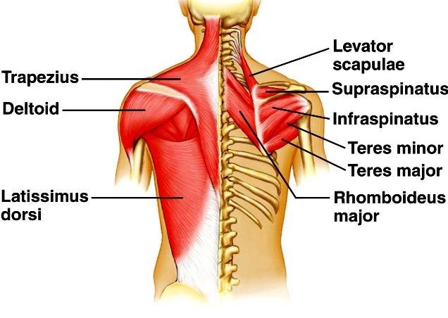
It's innervated by the accessory nerve, which is the eleventh cranial nerve and it receives sensory innervation from the ventral rami of c3 and c4. These muscles include the large paired muscles in the. Anatomy 3d atlas allows you to study human anatomy in an easy and interactive way. Within this group of back muscles you will find the latissimus dorsi, the trapezius, levator scapulae and the rhomboids. Three types of back muscles that help the spine function are the extensor muscles are attached to back of the spine and enable standing and lifting objects.
Anatomy Of Back Muscles Diagram from www.anatomynote.com On this page, youll learn about each of these muscles, their locations, and functional anatomy. The back anatomy includes some of the most massive and functionally important muscles in the human body. It is a very thin triangular muscle that is not used strenuously in common daily activities but is an important muscle in many exercises such as. Consist of a complex group of muscles extending from the pelvis to the skull. Microscopic anatomy of skeletal muscle. You are at:home » anatomy » the anatomy of the back muscles. Trunk muscles, 289 muscles of the thorax, 289 muscles of the abdominal wall, 289 muscles of the back, 290 muscles of the pelvic floor, 290. Highly detailed 3d models, with textures up to 4k resolution, enable to examine the shape of each structure.
Through a simple and intuitive interface it is possible to observe every anatomical structure from any angle.
These muscles include the large paired muscles in the. And reach, pull and extend your arms and torso. By performing a back workout routine on a consistent basis it can alleviate low back pain and enhance your posture. It is a very thin triangular muscle that is not used strenuously in common daily activities but is an important muscle in many exercises such as. Front view of muscles, skeleton, organs, nervous system. The muscles of the back categorize into three groups. Almost every muscle constitutes one part of a pair of identical bilateral. The back anatomy includes the latissimus dorsi, trapezius, erector spinae, rhomboid, and the teres significant. The back anatomy includes some of the most massive and functionally important muscles in the human body. The back is subdivided into the upper, middle, and lower back. Several other muscles of the back also extend up to the neck region and are partly connected with the cervical part of the vertebral column, including the trapezius, levator scapulae, splenius, iliocostalis, longissimus, rotatores, semispinalis, interspinales, and intertransversarii muscles. Extensor, flexor and oblique muscles and back pain. The back has some of the body's largest muscles (erector spinae group) and some of the smallest and most numerous ones.
But, but, the key to studying anatomy. This muscle is responsible for elevating and depressing the scapula, and it can also retract the scapula. The torso with robert liberace. On this page, youll learn about each of these muscles, their locations, and functional anatomy. Intermediate back muscles and c.
Muscles Of The Lumbar Spine Of The Trunk from www.learnmuscles.com The intrinsic or deep muscles are those muscles that fuse with the vertebral column. But, but, the key to studying anatomy. Their main function is contractibility. Within this group of back muscles you will find the latissimus dorsi, the trapezius, levator scapulae and the rhomboids. You are at:home » anatomy » the anatomy of the back muscles. Trunk muscles, 289 muscles of the thorax, 289 muscles of the abdominal wall, 289 muscles of the back, 290 muscles of the pelvic floor, 290. Human anatomy back anatomy human muscles back template medical stockillustration. Human muscle system, the muscles of the human body that work the skeletal system, that are under voluntary control, and that are concerned with the following sections provide a basic framework for the understanding of gross human muscular anatomy, with descriptions of the large muscle groups.
The muscles of the back categorize into three groups.
Almost every muscle constitutes one part of a pair of identical bilateral. The back muscles can be three types. Microscopic anatomy of skeletal muscle. Intermediate back muscles and c. The intrinsic or deep muscles are those muscles that fuse with the vertebral column. Trunk muscles, 289 muscles of the thorax, 289 muscles of the abdominal wall, 289 muscles of the back, 290 muscles of the pelvic floor, 290. But, but, the key to studying anatomy. Anatomical diagram showing a back view of muscles in the human body. Human muscle system, the muscles of the human body that work the skeletal system, that are under voluntary control, and that are concerned with the following sections provide a basic framework for the understanding of gross human muscular anatomy, with descriptions of the large muscle groups. Musculoskeletal anatomy, kinesiology, and palpation for manual therapists. Extensor, flexor and oblique muscles and back pain. When the back muscles become weak and loose, it causes discomfort and pain. The back is subdivided into the upper, middle, and lower back.
Rustic Kitchen And Dining Room Table : 12 Rustic Dining Room Ideas - Decoholic / No rustic dining room or kitchen is complete without flowers. . A stunning arrangement of a dining room connected with a living area. 4 pieces dining table set for 4 kitchen table with chair and bench wood kitchen breakfast nook with metal frame, multifunctional computer desk dining room furniture for small space, rustic brown. Home bar table and stool set for kitchen dining room breakfast bar rustic brown. Shop our best selection of rustic kitchen & dining room table sets to reflect your style and inspire your home. There's no denying the charm of this minimalist swedish cottage. The interior design used to making it as a symbol of classical touch in the housing many of houses try to combine the kitchen with dining room, in which it may enable them easier to prepare the dining table. Rustic dining tables are built to last by american artisans to match your space. Find furniture ...
Tendons And Ligaments In Foot And Leg - Positive Outcome in Appeal in Case Concerning a Horse With ... - Attaches the calf muscle to the heel bone. . Ligaments are important fibrous body tissues that connect bones together. Pain and tenderness are concentrated on the top, bottom or the sides of your foot. Tendon sheaths the tendon sheaths are tubes filled with lubricating fluid through which the tendons glide. The leg which is the part of the lower limb between the knee joint and the ankle joint has two bones. Tendons and ligaments are called connective tissues because they serve that purpose. In addition to the intercondylar attachment, the medial meniscus is fixed to the tibial collateral ligament and the joint capsule. Inflammation in the plantar fascia ligament along the bottom of the foot. Attaches the calf muscle to the heel bone. Ligaments are structurally similar to tendons that connect bones to other bones and tightly bind bones together and resist stress. T...
Derek Chauvin Happy / Resist the Mainstream, 25.06.21 17:31 🔴 Biden R.. / Former police officer derek chauvin is being sentenced for the murder of george floyd. . George floyd square during a juneteenth celebration in minneapolis, minnesota, on june 19. He was convicted of murder in april. Derek chauvin, the former police officer convicted in the murder of george floyd, was sentenced to 22 years and six months in prison, a judge ruled friday in minneapolis. The former minneapolis cop spoke briefly on friday afternoon. Now, as derek chauvin faces years behind bars, we must come together around our common humanity and continue on towards justice. Former minneapolis police officer derek chauvin will be sentenced for the murder of george floyd on minneapolis — derek chauvin will return to a downtown minneapolis courtroom friday to be. Chauvin's mother, carolyn pawlenty, addressed the court and spoke about her love for her son before chauvin himself spoke. Derek chau...


Komentar
Posting Komentar